
Blog
- Home
- /
- Blog
What Are the 2026 Top Types of Recombinant Collagen?
Recombinant Collagen is moving from a niche biotechnology topic into practical skincare, wound care, biomaterials, and laboratory research. In 2026, the leading types will not be judged by novelty alone. Their performance will depend on amino-acid sequence, triple-helix stability, purity, scalability, and documented biological response. Type I remains the best-known candidate for structural support. Type III may attract attention for softer tissue applications and skin-focused formulations. Type XVII is also gaining interest because of its relationship with epithelial and hair-follicle biology. These categories are not interchangeable.
Professor David L. Kaplan, a widely recognized biomaterials researcher, has described collagen as “a remarkable material because of its structural hierarchy.” That observation matters. A small vial of recombinant protein can hide major differences in folding, cross-linking, endotoxin levels, and degradation behavior. Yeast, bacteria, and mammalian cells can each produce useful collagen platforms. They do not create identical products. Manufacturing conditions leave fingerprints.
This guide compares the most discussed recombinant collagen types for 2026. It considers source systems, functional strengths, limitations, and evidence quality. Readers should examine real data, not polished claims. Some promising products still lack long-term clinical evidence. That weakness deserves attention. The “top” choice may change with the application, budget, and regulatory pathway. There is no universal winner. A transparent comparison is more useful than a confident ranking.
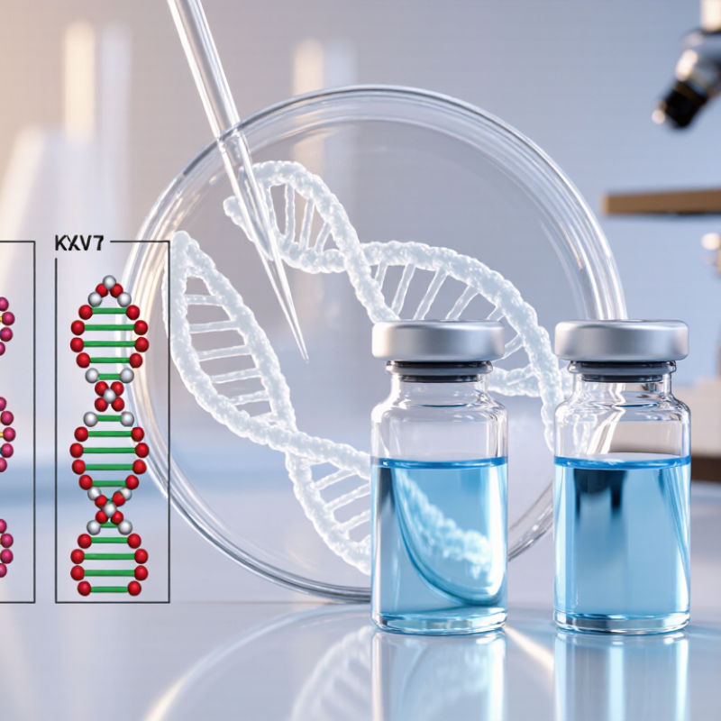
What Recombinant Collagen Is and Why Its Types Matter
What is recombinant collagen? It is collagen produced through controlled biological systems rather than direct extraction from animal tissue. Scientists place collagen-related genetic instructions into suitable host cells. These cells then produce a purified protein or collagen-like material. The final structure may resemble human collagen closely, but it is not always identical. That difference matters.
The most discussed recombinant collagen types in 2026 include Types I, III, II, and V. Type I is widely studied for skin, bone, and tendon applications.
Type III often appears in skin and soft-tissue research. Type II is associated with cartilage-focused materials. Type V can influence collagen-fiber organization.
Some products use full-length collagen, while others use shorter collagen peptides or collagen-like sequences. The “best” type depends on the intended tissue and formulation.
Type selection affects strength, hydration, cell interaction, and material stability. In practical development, researchers also examine molecular size, triple-helix formation, purity, and endotoxin levels. A clear gel, for example, may look impressive in a laboratory vial but perform poorly after storage.
The label recombinant alone proves little. Independent testing remains important.
I also think simplified type charts can mislead, because real tissues contain several collagen types together. More testing is still needed for long-term performance, especially when laboratory results are transferred to human use.
How Recombinant Collagen Types Are Classified in 2026
In 2026, recombinant collagen is best classified by what its sequence is designed to represent, rather than by one universal ranking. Type I is associated with skin, tendon, and bone; type III is common in skin and blood vessels; type II is linked to cartilage. These labels describe biological models, not automatic proof that a product behaves exactly like collagen in human tissue.
A second useful distinction is molecular form. Some materials contain collagen-like triple-helical regions, while others use shorter peptides or modified sequences. Check whether a supplier reports the sequence, molecular size, and evidence for its structure. Small details matter. A clear vial does not reveal how closely its contents resemble native collagen.
Researchers also classify materials by production host and intended use, such as laboratory studies, biomaterials, or topical formulations. The host can affect processing and consistency, but it does not determine quality on its own. In practice, compare composition, testing methods, and stated limitations. The categories can blur: engineered collagen may combine features from different natural types. That makes simple “top type” lists tempting, but not very informative. A careful classification should say what was measured and what remains uncertain.
What Are the 2026 Top Types of Recombinant Collagen? - How Recombinant Collagen Types Are Classified in 2026
A science-based comparison of the principal recombinant collagen types used or investigated for biomaterials, tissue engineering, wound care, and research applications.
| Collagen Type | Structural Class | Primary Native Location | Key Biological Function | Typical Recombinant Product Format | Common Application Areas | Important Development Considerations |
|---|---|---|---|---|---|---|
| Type I | Fibrillar | Skin, bone, tendon, ligament, dentin, and many connective tissues | Provides high tensile strength and forms the major structural framework of many tissues | Full-length or functional fragments; soluble recombinant protein; engineered collagen-like polymers | Wound-care matrices, tissue scaffolds, cell culture substrates, bone-related biomaterials, and research reagents | Native triple-helix formation, thermal stability, fibril assembly, and control of immunogenic impurities are important |
| Type II | Fibrillar | Articular cartilage, vitreous body, and other cartilage-rich tissues | Supports cartilage tensile integrity and interacts with proteoglycans within the extracellular matrix | Recombinant triple-helical domains, full-length constructs, or collagen-mimetic sequences | Cartilage research, chondrocyte culture, osteochondral models, and musculoskeletal tissue engineering | Correct folding, resistance to enzymatic degradation, and preservation of cartilage-relevant cell signaling are key challenges |
| Type III | Fibrillar | Skin, blood-vessel walls, intestinal tissue, uterus, and other extensible organs | Contributes elasticity and forms fibrils together with Type I collagen | Soluble recombinant collagen, collagen-like domains, or Type I/III-inspired composite materials | Dermal repair, soft-tissue scaffolds, vascular models, wound healing, and cell-growth substrates | Fibril co-assembly, mechanical compliance, degradation rate, and maintenance of a physiologically relevant Type I/III balance require control |
| Type V | Fibril-regulating | Interspersed with Type I collagen in skin, cornea, bone, and other connective tissues | Regulates collagen fibril nucleation, diameter, and organization | Recombinant chain fragments, collagen-binding domains, or engineered regulatory peptides | Fibrillogenesis studies, corneal models, composite scaffolds, and extracellular-matrix research | It is generally a minor fibrillar component; biological performance depends strongly on its ratio and interaction with Type I collagen |
| Type IV | Network-forming | Basement membranes beneath epithelia and around blood vessels | Forms sheet-like networks that provide filtration, structural support, and cell-adhesion signals | Recombinant non-collagenous domains, selected network-forming fragments, or engineered matrix proteins | Basement-membrane models, organoids, barrier studies, cell adhesion, and regenerative medicine research | Type IV collagen contains multiple genetically distinct alpha chains; accurate network assembly and domain-specific function are technically demanding |
| Type VII | Anchoring fibril-forming | Dermal–epidermal junction of the skin and related epithelial basement membranes | Anchors the basement membrane to the underlying dermal matrix and helps maintain tissue cohesion | Recombinant anchoring domains, functional fragments, or engineered adhesion proteins | Skin-barrier models, epithelial tissue engineering, wound repair research, and adhesion assays | Its very large size, specialized terminal domains, and anchoring-fibril assembly make full-length recombinant production difficult |
| Type XVII | Transmembrane | Hemidesmosomes at the basal surface of stratified epithelial cells | Links intracellular adhesion structures with the basement membrane and supports epithelial attachment | Recombinant extracellular domains, binding fragments, or domain-specific research proteins | Skin biology, epithelial adhesion studies, hemidesmosome research, and disease-model development | It is a membrane-associated collagen with a large non-collagenous region; maintaining native domain orientation and proteolytic processing is important |
Classification basis: Recombinant collagen is commonly classified by native collagen type, structural organization, biological role, engineered sequence, and production format. In 2026, Type I, II, and III remain the principal fibrillar reference types, while Types IV, V, VII, and XVII represent important specialized categories for basement-membrane, fibril-regulation, epithelial-adhesion, and tissue-interface applications. “Top” indicates high scientific and development relevance rather than a company-specific market ranking.
Type I Recombinant Collagen: Features and Main Applications
Type I recombinant collagen is one of the most studied collagen formats for 2026 biomaterial development. It is produced by engineered microorganisms or cultured cells, rather than extracted directly from animal tissue. This can improve batch consistency and reduce concerns linked to source variability. Its amino acid sequence supports a stable triple-helix structure when processing conditions are carefully controlled. Small changes in temperature, pH, or purification can still affect performance.
In tissue engineering, Type I recombinant collagen can form porous scaffolds for skin, bone, tendon, and cartilage research. A dry scaffold may feel light, yet absorb liquid quickly and create a moist surface for cell attachment. Researchers also use it in hydrogels, coatings, and three-dimensional culture systems. Wound-care materials may benefit from its biological familiarity, but clinical performance depends on crosslinking, sterility, degradation rate, and mechanical strength. Cosmetic formulations may use hydrolyzed forms, although topical collagen should not be confused with implanted collagen.
The strongest advantage is control. It may offer defined composition and easier process tracking. However, “recombinant” does not automatically mean superior. Some products show limited strength or poor stability without additional processing. I have found that application-specific testing matters more than attractive specification sheets. Cell compatibility, endotoxin levels, residual host-cell proteins, and storage behavior require documented evaluation. Results from a laboratory scaffold may also change after sterilization or long-term hydration. That gap deserves more attention.
Types III, IV, and V Recombinant Collagen Compared
Type III, IV, and V recombinant collagen serve different biological roles. Type III commonly appears beside Type I in skin, blood vessels, and flexible connective tissue. It may support studies involving elasticity, wound repair, and matrix remodeling. Type IV forms sheet-like basement membranes beneath epithelial cells. Researchers often examine it in barrier models, kidney studies, and cell adhesion experiments. Type V helps regulate fibril formation, especially alongside Types I and III. Small changes may influence fibril diameter and tissue organization.
The comparison is not perfectly clean. Recombinant production systems can alter folding, hydroxylation, chain assembly, and final bioactivity. A Type IV material may look suitable on paper, yet perform poorly in a three-dimensional barrier model. Type III may offer better relevance for dermal research, while Type V can be more useful when fibril structure matters. Test the actual material.
Quality evidence should include sequence confirmation, purity data, endotoxin testing, and a relevant functional assay. Solubility also matters. A clear solution at low temperature may prevent unwanted aggregation during handling. I would compare materials under identical concentrations, buffers, and incubation times. Otherwise, the experiment measures preparation differences rather than collagen biology. The best choice depends on the model, not on a simple ranking. Even experienced teams can overinterpret an attractive specification sheet.
How to Select the Right Recombinant Collagen Type for Each Use
What Are the 2026 Top Types of Recombinant Collagen?
How to Select the Right Recombinant Collagen Type for Each Use
Type I remains the main choice for skin matrices, wound research, and bone-related scaffolds. It offers strong fibril formation and useful structural support. Type III is often paired with Type I when softer, more elastic tissue behavior matters. This combination can suit dermal models and vascular research. Type II is more specialized. It fits cartilage studies, joint-focused formulations, and chondrocyte culture. Type XVII is gaining attention for epithelial and basement membrane research, though its use remains more selective.
The collagen type should follow the tissue target, not market popularity. Check the amino acid sequence, molecular size, triple-helix stability, and fibril-forming behavior. For cell culture, low endotoxin levels and consistent lot performance are essential. For injectable or implant-related research, assess purity, residual host-cell proteins, bioburden controls, and available technical documentation. Human-sequence recombinant collagen may reduce some source-related concerns, but it does not remove every biological risk.
A practical screening table helps. Match Type I with tensile support, Type II with cartilage signals, and Type III with elasticity. Then test concentration, crosslinking, and degradation rate. Small changes can alter cell attachment dramatically. No type wins every time. The awkward part is that published data may not predict performance in your exact matrix. Run a small pilot study before scaling. Recheck the choice when the scaffold becomes thicker, stiffer, or more highly hydrated.